|
|
< CNMD \ <Tutorial \<Second step \ 7-point checklist
|
 |
|
7-Point Checklist
[Argenziano et al. Arch Dermatol 1998]
|
|
Additional diagnostic algorithm developed for simplifying the classic pattern analysis
-
Low number of features to identify
-
Scoring diagnostic system
|
 |
Consensus Meeting (1990)
|
|
7-Point Checklist (1998)
|
-
Discrete Pigment Network
-
Prominent PN
-
Regular PN
-
Irregular PN
-
Wide PN
-
Narrow PN
-
Broad PN
-
Delicate PN
|
 |
1. Atypical Pigment Network |
-
Pseudopods
-
Radial streaming
|
|
2. Irregular Streaks |
-
Brown globules
-
Black dots
|
|
3. Irregular Dots/globules |
- Whitish veil
|
|
4. Blue - whitish veil |
- White scarlike areas
- Gray - blue areas
|
|
5. Regression Structures |
- Hypopigmented areas
(not siginificant)
- Reticular depigmentation
(not significant)
|
|
(ADDITIONAL CRITERIA) |
- Milia-like cysts
- Comedo-like openings
- Telangiectasia
- Red-blue areas
- Maple leaf-like areas
|
 |
Criteria for
non-melanocytic
lesions
|
6. Irregular Pigmentation
7. Atypical Vascular Pattern |
|
|
7-Point Scored Diagnosis
|
-
Odds ratios of each of the 7 criteria were calculated by multivariate analysis.
-
7-point score of 2 was given to the 3 criteria with odds ratios more than 5, and a score of 1 was given to the 4 criteria with odds ratios less than 5.
-
By simple addition of the individual scores a minimum total score of 3 is required for the diagnosis of melanoma, whereas a total score of less than 3 is indicative of a non-melanoma.
|
7-Point Checklist: Definition and Histopathologic Correlates of the 7 Melanoma-Specific Dermoscopic Criteria
|
|
Criterion
|
Definition
|
Histopathologic correlates
|
1. Atypical pigment network
|
Black, brown, or gray network with irregular meshes and thick lines |
Irregular and broadened rete ridges |
2. Blue-whitish veil
|
Irregular, confluent, gray-blue to whitish-blue diffuse pigmentation |
Acanthotic epidermis with focal hypergranulosis above sheets of heavily pigmented melanocytes in the dermis |
3. Atypical vascular pattern
|
Linear-irregular or dotted vessels not clearly combined with regression structures |
Neovascularization |
4. Irregular streaks
|
Irregular, more or less confluent, linear structures not clearly combined with pigment network lines |
Confluent junctional nests of melanocytes |
5. Irregular pigmentation
|
Black, brown, and/or gray pigmented areas with irregular shape and/or distribution
|
Hyperpigmentation throughout the epidermis and/or upper dermis
|
6. Irregular dots/globules
|
Black, brown, and/or gray round to oval, variously sized structures irregularly distributed within the lesion
|
Pigment aggregates within stratum corneum, epidermis, dermo-epidermal junction, or papillary dermis
|
7. Regression structures
|
White areas (white scarlike areas) and blue areas (gray-blue areas, peppering, multiple blue-gray dots) may be associated, thus featuring so-called blue-whitish areas virtually indistinguishable from blue-whitish veil |
Thickned papillary dermis with fibrosis and/or variable amounts of melanophages
|
|
|
7-Point Checklist. A minimum total score of 3 is required for the diagnosis of melanoma
|
|
ELM criterion
|
Odds ratioa
|
7-point scoreb
|
Major criteria:
1. Atypical pigment network |
5.19 |
2 |
| 2. Blue-whitish veil |
11.1 |
2 |
| 3. Atypical vascular pattern |
7.42 |
2 |
Minor criteria:
4. Irregular streaks |
3.01 |
1 |
| 5. Irregular pigmentation |
4.90 |
1 |
| 6. Irregular dots/globules |
2.93 |
1 |
| 7. Regression structures |
3.89 |
1 |
| a Odds ratios measuring the capacity of each criterion to increase the probability of melanoma diagnosis. |
| b The score for a given criterion is determined on the basis of the odds ratio: >5 (score = 2), and <5 (score = 1). |
|
|
Simply add the scores of each criterion that is present within a pigmented lesion.
|
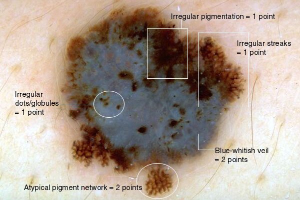
Melanoma:
7point score = 7
|
|
Melanoma:
7point score = 3
|
|
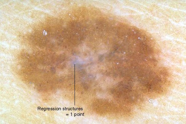
Clark nevus:
7point score = 1
|
|
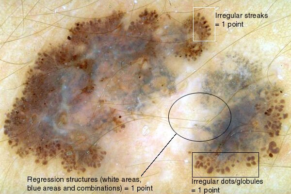
Melanoma:
7point score = 6
|

|
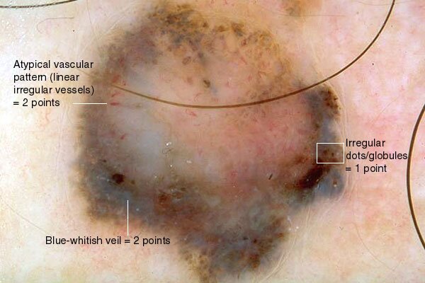
Melanoma:
7point score = 5
|
|
DS Medica S.r.l. | CF/P.IVA 12676030153 | © DS Medica S.r.l. | Privacy | Contatti 
|