1 - Milia-like cysts
2 - Comedo-like openings
3 - Exophytic papillary structures
4 - Red Lacunas
5 - Leaf-like areas
6 - Central White patch
Milia-like cysts
This typical example of seborrheic keratosis is characterized by numerous milia-like cysts (arrows) and few comedo-like openings
|
|
)
|
)
|
|
|
)
|
)
|
|
|
)
|
|
Comedo-like openings
This heavily pigmented seborrheic keratosis is characterized by numerous comedo-like openings (arrows). Only few milia-like cysts can be recognized.
|
|
)
|
)
|
|
|
)
|
)
|
|
|
)
|
)
|
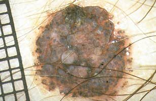
Exophytic papillary structures
This papillomatous dermal nevus (Unna nevus) is characterized by several, closely aggregated exophytic papillary structures. Nearly identical papillations are commonly found also in seborrheic keratoses.
|
|
)
|
 |
|
|
 |
)
|
|
|
)
|
|
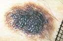
Red Lacunas
Stereotypical example of hemangioma with numerous red-bluish lacunas
|
|
)
|
)
|
|
|
)
|
)
|
|
|
)
|
|
Leaf-like areas
The clue for the diagnosis of this basal cell carcinoma is the presence of leaf-like areas (arrows) at the periphery of this lesion. Obviously one needs to have some visual imagination to recognize leaf-like structures!!
|
|
|
|
|
|
|
|
|
|
|
|
)
|
|
)
)
)
)
Central White patch
The central white patch represents the dermoscopic hallmark of dermatofibroma, as illustrated in this example
|
|
|
|
|
|
|
|
|
|
)
)
)
)